Top Medical Imaging Technologies in 2024

Introduction
Medical imaging technologies have advanced rapidly over the past few decades, playing a critical role in the diagnosis and treatment of various health conditions. As we step into 2024, the landscape of medical imaging continues to evolve, driven by technological innovations that enhance precision, reduce risks, and improve patient outcomes. This article delves into the top medical imaging technologies of 2024, exploring the latest advancements and their impact on healthcare.
Magnetic Resonance Imaging (MRI): The Gold Standard
Magnetic Resonance Imaging (MRI) remains one of the most powerful tools in medical imaging. In 2024, MRI technology has seen significant enhancements, particularly in the areas of speed, resolution, and functional imaging.
High-resolution MRI has become more accessible, providing detailed images of soft tissues, including the brain, muscles, and internal organs. This level of detail is crucial for diagnosing conditions such as tumors, strokes, and musculoskeletal disorders.
Moreover, functional MRI (fMRI) has advanced, allowing for more precise mapping of brain activity. This is especially important in neurology and psychiatry, where understanding brain function can lead to better treatments for conditions like epilepsy, depression, and anxiety disorders. The integration of AI in MRI systems has further improved image analysis, making it faster and more accurate, thereby enhancing diagnostic capabilities.
Computed Tomography (CT) Scans: Faster and More Accurate
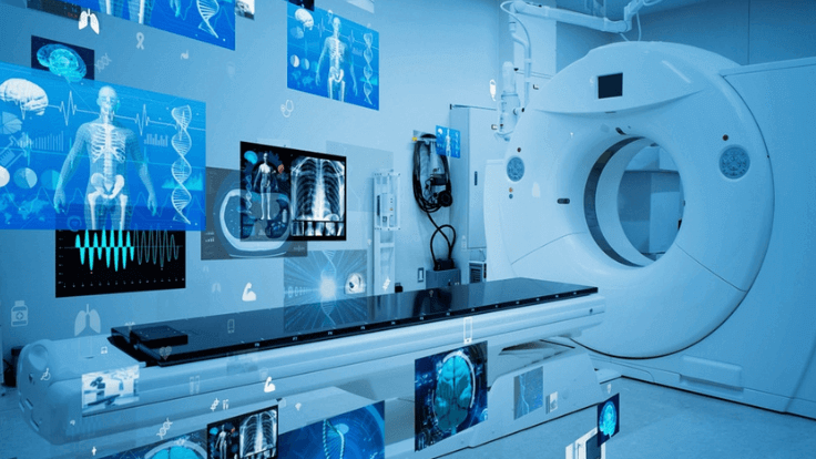
Computed Tomography (CT) scans have long been a staple in medical imaging, particularly for their ability to provide detailed images of the body’s internal structures. In 2024, CT technology has evolved to offer even faster imaging with lower radiation doses, making it safer for patients.
Dual-energy CT scanners, which use two different energy levels of X-rays, have gained prominence. These scanners can differentiate between materials more effectively, improving the accuracy of diagnoses for conditions like kidney stones, vascular diseases, and tumors. Additionally, the speed of modern CT scanners has drastically reduced the time required for full-body scans, enhancing patient comfort and throughput in busy clinical settings.
AI-powered image reconstruction algorithms now enable clearer images with less noise, even in low-dose scans. This innovation is particularly beneficial in pediatric imaging and other sensitive cases where minimizing radiation exposure is crucial.
Ultrasound: From 2D to 3D and Beyond
Ultrasound technology has advanced significantly in 2024, moving beyond traditional 2D imaging to offer 3D, 4D, and even Doppler ultrasound capabilities. These advancements have expanded the use of ultrasound in areas such as obstetrics, cardiology, and emergency medicine.
3D and 4D ultrasounds provide more detailed and dynamic views of the fetus during pregnancy, allowing for better monitoring of development and early detection of abnormalities. In cardiology, 4D echocardiography offers real-time imaging of the heart, improving the diagnosis of congenital heart diseases and valve disorders.
Portable ultrasound devices have also become more widespread, making it easier for healthcare providers to perform scans at the bedside or in remote locations. These devices are particularly valuable in emergency situations where quick and accurate diagnosis is critical. AI integration in ultrasound is improving image interpretation, reducing the need for highly specialized operators, and making the technology more accessible in a variety of healthcare settings.
Positron Emission Tomography (PET) Scans: Enhanced Precision in Oncology
Positron Emission Tomography (PET) scans have become increasingly important in oncology, providing detailed images of metabolic activity within the body. In 2024, advancements in PET technology have further enhanced its precision and utility in cancer diagnosis and treatment.
Hybrid PET/CT and PET/MRI scanners combine the strengths of both modalities, offering detailed anatomical and functional images in a single session. This combination allows for more accurate staging of cancers, better assessment of treatment response, and improved planning for radiation therapy.
Moreover, new radiotracers have been developed that target specific types of cancer cells, improving the detection of early-stage tumors and metastases. These advancements have made PET scans an indispensable tool in personalized medicine, where treatments are tailored to the individual characteristics of a patient’s cancer.
Artificial Intelligence (AI) in Medical Imaging: Revolutionizing Diagnostics
Artificial Intelligence (AI) continues to revolutionize medical imaging in 2024, enhancing the speed, accuracy, and accessibility of diagnostics. AI algorithms are increasingly used to analyze images from various modalities, including MRI, CT, and ultrasound, identifying patterns that may be missed by the human eye.
AI-powered diagnostic tools can process vast amounts of data quickly, providing real-time analysis and decision support to radiologists. This not only speeds up the diagnostic process but also reduces the likelihood of human error. In some cases, AI systems have been shown to match or even surpass human experts in detecting certain conditions, such as lung cancer on chest X-rays or diabetic retinopathy in eye scans.
Furthermore, AI is playing a crucial role in image enhancement and reconstruction, enabling clearer images from lower-quality scans. This is particularly beneficial in resource-limited settings, where access to high-end imaging equipment may be restricted.
Advanced Fluoroscopy: Real-Time Imaging with Lower Doses
Fluoroscopy, a technique that provides real-time moving images of the inside of the body, has seen significant advancements in 2024. The latest fluoroscopy systems offer higher resolution images with lower radiation doses, making procedures safer for both patients and healthcare providers.
These advancements are particularly beneficial in interventional radiology, where fluoroscopy is used to guide minimally invasive procedures such as angiography, stent placement, and catheter insertion. The improved image quality allows for more precise interventions, reducing the risk of complications.
Additionally, the development of portable fluoroscopy units has expanded the use of this technology in settings such as emergency rooms and operating theaters, where immediate imaging is critical.
Dual-Energy X-ray Absorptiometry (DEXA): Beyond Bone Density
Dual-Energy X-ray Absorptiometry (DEXA) has traditionally been used to measure bone density and diagnose osteoporosis. In 2024, DEXA technology has evolved to offer more comprehensive body composition analysis, including the measurement of fat and lean muscle mass.
This expanded capability is particularly useful in fields such as sports medicine, where precise measurement of body composition can aid in the optimization of training programs and injury prevention. In clinical settings, DEXA scans are increasingly used to assess the risk of fractures in patients with metabolic disorders, as well as to monitor the effectiveness of treatments for conditions like obesity and sarcopenia (age-related muscle loss).
The latest DEXA machines offer faster scans with lower radiation doses, making them safer and more comfortable for patients. The integration of AI in DEXA analysis has also improved the accuracy and reproducibility of results, enhancing the clinical utility of this technology.
Optical Coherence Tomography (OCT): Precision in Ophthalmology
Optical Coherence Tomography (OCT) is a non-invasive imaging technique that provides high-resolution cross-sectional images of the retina. In 2024, OCT technology has continued to advance, offering even greater precision in diagnosing and monitoring eye conditions.
The latest OCT systems provide enhanced imaging of the retinal layers, allowing for earlier detection of conditions such as glaucoma, macular degeneration, and diabetic retinopathy. These advancements have improved the ability of ophthalmologists to monitor disease progression and tailor treatments to the specific needs of each patient.
Furthermore, the introduction of portable OCT devices has made this technology more accessible in a variety of healthcare settings, including primary care offices and remote clinics. This increased accessibility is helping to improve the early diagnosis and management of eye diseases, ultimately reducing the risk of vision loss.
Molecular Imaging: A New Frontier in Personalized Medicine
Molecular imaging is an emerging field that involves the visualization of cellular and molecular processes within the body. In 2024, molecular imaging technologies are making significant strides, offering new insights into the underlying mechanisms of diseases and enabling more personalized approaches to treatment.
Techniques such as Single Photon Emission Computed Tomography (SPECT) and hybrid PET/MRI are at the forefront of molecular imaging, allowing for the visualization of specific biological processes in real-time. These technologies are particularly valuable in oncology, where they can be used to assess the effectiveness of targeted therapies and monitor the response to treatment at the molecular level.
The ability to visualize molecular changes in tissues before structural changes become apparent has the potential to revolutionize early diagnosis and treatment planning. As molecular imaging continues to evolve, it is expected to play an increasingly important role in personalized medicine, enabling treatments that are tailored to the unique characteristics of each patient’s disease.
Theranostics: Combining Imaging and Therapy
Theranostics is a cutting-edge field that combines diagnostic imaging with targeted therapy, offering a more integrated approach to treating diseases such as cancer. In 2024, theranostics is gaining traction as a promising strategy for improving treatment outcomes and reducing side effects.
The concept of theranostics involves using imaging techniques, such as PET or SPECT, to identify specific biomarkers associated with a disease. Once identified, targeted therapies, often radiopharmaceuticals, are administered to treat the disease at its source. The effectiveness of the therapy is then monitored using the same imaging techniques, allowing for real-time adjustments to the treatment plan.
Theranostics has shown particular promise in the treatment of cancers, such as prostate cancer and neuroendocrine tumors, where traditional therapies may be less effective. By combining diagnosis and treatment in a single approach, theranostics offers the potential for more precise and effective care, tailored to the individual characteristics of the patient’s disease.
Conclusion
The field of medical imaging continues to evolve rapidly, with 2024 marking another year of significant advancements. From enhanced MRI and CT scans to the integration of AI and the rise of molecular imaging, these technologies are revolutionizing the way healthcare providers diagnose and treat a wide range of conditions. As these innovations continue to develop, they hold the promise of improving patient outcomes, reducing risks, and enabling more personalized approaches to healthcare. Staying informed about the latest trends in medical imaging is essential for healthcare professionals and patients alike, as these technologies will undoubtedly shape the future of medicine.


